Biograph Study Finds Coronary Inflammation in Many Asymptomatic Adults
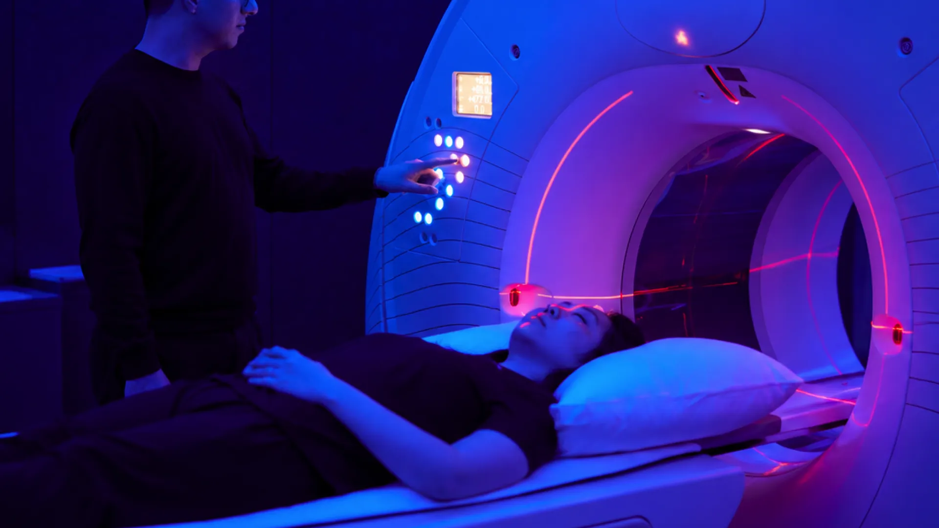
Biograph has reported that 42.9% of asymptomatic, low-risk adult participants in its first U.S. study of Caristo Diagnostics' CaRi-Heart technology showed elevated coronary inflammation. The results were published on October 22, 2026.
According to Biograph, 17.9% of participants had very high inflammation in at least one major heart artery. Among participants with no detectable coronary plaque, the fatty buildup inside arteries, about 47% still exhibited elevated coronary perivascular inflammation, meaning inflammation in the fat tissue surrounding the vessel. Plaque-free arteries, in other words, did not rule out the inflammatory signal the study was designed to detect.
The study combined high-resolution coronary computed tomography angiography (CCTA), a CT scan that images the heart's arteries, with cardiometabolic profiling, a review of metabolic markers such as cholesterol and blood sugar. Image analysis was performed with Caristo's CaRi-Heart and CaRi-Plaque technologies. Participants were clinically eligible members of Biograph's Black tier, the clinic's premium membership level, who were offered the study.
The conventional markers did not track the imaging finding. Per Biograph, ApoB (a protein that carries LDL cholesterol particles), non-HDL-C, HbA1c (a roughly three-month average of blood glucose), high-sensitivity CRP (a blood test for systemic inflammation) and the AHA PREVENT-ASCVD risk calculator showed no correlation with the presence of coronary inflammation. That is a notable result for clinicians who rely on lipid and risk-score panels to decide who needs closer evaluation.
The study has a defined history. Biograph and Caristo announced it on September 30, 2025, describing it as a first-of-its-kind U.S. study to advance AI-powered prevention. Biograph describes itself as the first U.S. clinic to study CaRi-Heart for early detection of coronary artery disease in patients without symptoms, a claim stated on Caristo's preventive screening page. Caristo describes CaRi-Heart as the first and only biomarker to measure coronary inflammation and assess cardiovascular mortality risk, and characterizes CaRi-Heart Analysis as an AI-enabled imaging solution that quantifies coronary inflammation and characterizes plaque.
On the regulatory side, CaRi-Heart has received FDA De Novo marketing authorization to quantify coronary inflammation from routine coronary CT angiography. CaRi-Plaque has received FDA clearance for AI-assisted coronary plaque and stenosis analysis. The distinction matters for anyone evaluating deployment. The inflammation measurement is derived from images that a patient already undergoes for a CCTA, so it does not require a separate acquisition.
In my view, the central question for this dataset is what the number describes. The participants were a self-selected group of clinic members, not a general population sample, so 42.9% is a prevalence within that cohort and should not be read as a national estimate. The 47% figure among plaque-free patients is more striking to me, because it suggests that the standard anatomical question, whether plaque is present, may miss a biological process that imaging can now quantify.
The broader context here is outcomes. Perivascular fat inflammation is a candidate marker, not an established endpoint, and the reported findings describe prevalence rather than subsequent cardiovascular events. Whether elevated coronary inflammation in asymptomatic adults predicts later events, and whether treating it changes those events, are questions that prospective follow-up and intervention trials would need to answer. Caristo's mortality-risk claim for CaRi-Heart rests on that kind of evidence, and a single site's cohort is a starting point for it.
Still, the practical direction is clear enough to name. If a quantitative inflammation readout can be generated from scans already being acquired, and if it captures risk that lipid panels and PREVENT scores do not, then prevention workflows could start earlier and rely less on the presence of symptoms or plaque. For clinicians and health-IT teams, the open work is integration: how the score is reported, who acts on it, and how it sits alongside existing risk tools. The study is an early, clearly bounded step in that direction, and its value will depend on what follow-up shows.